Evaluation of Fusion Following Anterior Cervical Discectomy and Fusion by Stand Alone PEEK Cage for Cervical Disc Herniation: Experience in a Tertiary Level Hospital in Bangladesh.

Volume 8 | Issue 2 | July-December 2023 | Page: 00-00 | Wayez Mahbub, Md. Anowarul Islam, Shagor Kumar Sarker, Suvradev Saha, Humaira Sultana

DOI: https://doi.org/10.13107/ijs.2023.v08.i02.00

Submitted: 29/08/2023; Reviewed: 11/09/2023; Accepted: 21/10/2022; Published: 10/12/2023


Authors: Wayez Mahbub [1], Md. Anowarul Islam [1], Shagor Kumar Sarker [1], Suvradev Saha [1], Humaira Sultana [1]

[1] Department of Orthopaedics and Spine surgery, Dhaka Community Medical College, Dhaka, Bangladesh.

Address of Correspondence

Dr. Wayez Mahbub
Registrar, Department of Orthopaedic and Spine Surgery, Dhaka Community Medical College, Dhaka, Bangladesh.
Email: wayez1134@gmail.com


Abstract

Background: Cervical disc herniation may result in direct compression of the spinal cord or impingement of the nerve roots. Different surgical procedures are performed in cervical disc herniation to relief cord compression, as well as achieving stabilization. Among the surgical techniques anterior cervical discectomy and fusion (ACDF) is the most commonly used technique. Various fixation and fusion cages are used in ACDF but Standalone PEEK cage is the standard one. To counteract the complications with the plating for ACDF, Stand-alone cage concept was constructed and favourable outcomes have been described. ACDF by stand-alone PEEK cage can provide immediate load bearing support to the anterior column and also facilitate fusion.

Materials & methods: This study is accomplished in the spine division, department of Orthopaedic Surgery, Bangabandhu Sheikh Mujib Medical University, Dhaka from January 2016 to January 2021. A total number of 70 patients with cervical disc herniation presenting with radiculopathy or myelopathy were included in this study. Patients with fractures, infections, tumours, or previous cervical spine surgery were excluded. Follow-up were done at 3 months, 6 months, 12 months and finally at 24 months post-operative. Bridwell’s criteria was used to assess radiological fusion.

Results: Mean patient age was 43.4 ± 6.24 years. Among them, 47 were men and 23 were women. 46 and 24 patients had one and two-level cervical disc surgeries, respectively. All of the study patients had neck pain and radiculopathy. 48 patients (68.6%) had myelopathic signs. Radiological outcome was assessed by Bridwell fusion criteria, radiological fusion was found in majority of patients after 6, 12 and 24 months follow up (71.42%, 80% and 90%, respectively) (p value <0.05).

Conclusion: ACDF by stand-alone PEEK cage is an excellent technique for the treatment symptomatic cervical disc herniation with significant fusion rate.

Keywords: Disc herniation, ACDF, Stand-alone PEEK cage


Introduction:

Disc herniation usually occurs when there is postero-lateral annular tear or stress and often accompanied with early stages of disc degeneration. [1]. Herniation at cervical level may ensue direct pressure over the spinal cord or may cause nerve root impingement. Cervical disc herniation may present with radiculopathy due to root compression or also with myelopathy due to cord compression. People after the fourth decade of life are affected more with male predilection. Male to female ratio is 1.4:1 [2]. For severe or progressive neurological compromise and significant non refractory pain requires operative treatment over conservative treatment as recommended by most guidelines [3]. Surgical procedures performed in cervical disc herniation are aimed at relieving spinal cord and root compression, as well as achieving stabilization. Among the surgical approaches and techniques, anterior cervical discectomy and fusion is the most preferred one [4]. This procedure is used to decompress the spinal cord and nerve root, to stabilize the affected segments and to restore intervertebral disc height [5]. Fusion rate following ACDF is approximately 92 % [6]. Different types of cages are used in ACDF surgery made up of titanium, PEEK cage, carbon fiber etc. [7]. ACDF by stand-alone PEEK cage provides immediate load bearing support to the anterior column and may facilitate fusion [6].

Materials and methods:
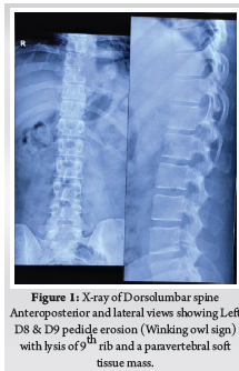
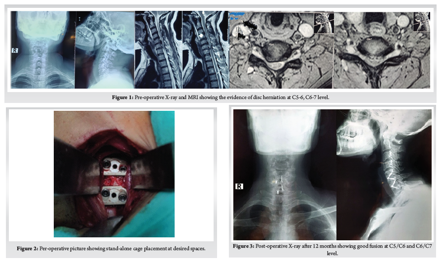
This Study was approved by the institutional review board of Bangabandhu Sheikh Mujib Medical University. This study was conducted in the spine division of Orthopaedic Surgery department of Bangabandhu Sheikh Mujib Medical University, Shahbag, Dhaka from January 2016 to January 2021. A total number of 70 patients with cervicaldisc herniation presenting with radiculopathy or myelopathy were included in this study. Patients with fractures, infections, tumours, or previous cervical spine surgery were excluded. Patients were followed up at 3 months, 6 months, 12 months and finally at 24 months. Bridwell’s criteria was used for radiological fusion.X-ray taken in A/P and Lateral view in every follow up to evaluate the fusion status. Fusion criteria as follow a) anterior-posterior bony bridging, b) bony continuity between upper and lower endplate, c) trabecular structure both anterior & posterior through the fenestration of PEEK cage, d) lack of radiolucent line around PEEK cage. (Fig. 1)

Statistical analysis:
Quantitative data analysis was done by Statistical Package for Social Science (SPSS-26). p<0.05 was regarded as the level of significance. Chi-Square test was used to assess and compare the categorical data.

Surgical procedure:
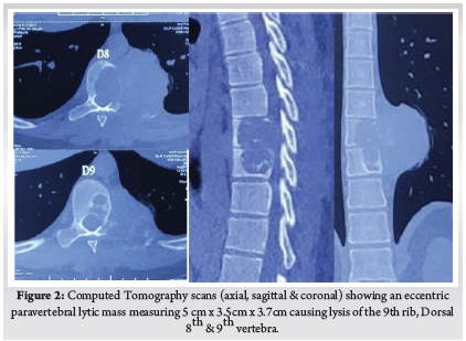
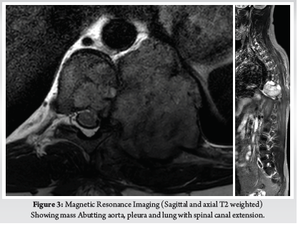
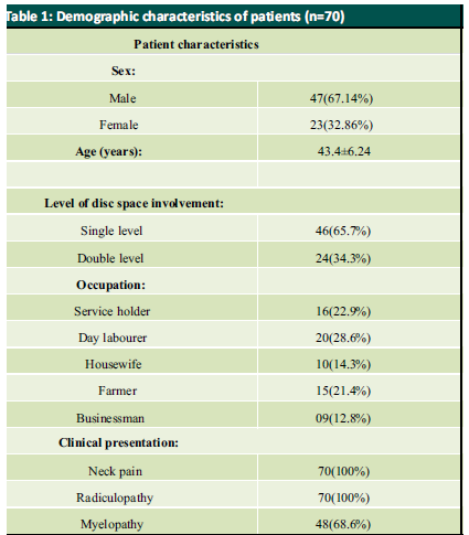
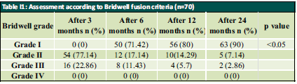
Patient was placed in supine position. The Gardener-wells tong traction was applied following anaesthesia. To keep the neck extended a sandbag was placed between shoulder blades. Patient’s head was rotated slightly to the opposite of the planned approach site. Then transverse incision was made. Platysma was identified and longitudinally incised. Anterior border of sternocleidomastoid was identified and then longitudinally incised the superficial layer of deep cervical fascia. Carotid pulse was localized by palpation. Then a cleavage was made between trachea, oesophagus medially and sternocleidomastoid with carotid sheath laterally. Then self-retaining retractor was placed accordingly. Prevertebral fascia was dissected and then longus colli muscle were retracted and elevated on both sides. To confirm the appropriate disc level, per-operative image intensifier was used following guide pin introduction. Then total discectomy was carried out followed by decortication of endplates by using curette. Lastly Stand-alone PEEK cage/ cages was inserted and fixed with screws (Fig. 2, 3). Again, image intensifier was used to check the final position of cage. Hemostasis was ensured by gel-foam and electrocautery. A drain tube was kept in situ. Platysma, subcutaneous layer and skin were closed accordingly. Before extubation cervical collar was used in every patient. Patients were encouraged to ambulate on the 5th post-operative day.

Results:
Mean patient age was 43.4 ± 6.24 years. Among them, 47 were men and 23 were women with a male: female ratio of 2:1. Patient demographic characteristics are illustrated in Table I. Within the sample, 46 and 24 patients had one and two-level surgeries, respectively. All of the study patients had neck pain and radiculopathy. 48 patients (68.6%) had myelopathic signs. Radiological outcome was assessed by Bridwell fusion criteria, radiological fusion was found in majority of patients after 6, 12 and 24 months follow up (71.42%, 80% and 90%, respectively) (p value <0.05) [Table II]


Discussion:
ACDF is considered to be the gold standard surgical technique for patients with cervical disc disease. Interbody fusion using tri-cortical bone graft provides good clinical outcome but there is worrisome donor site morbidity [8]. To combat this situation, use of different interbody fusion cages emerges like titanium, PEEK, Stand-alone cages etc. Though the fusion rate is quite similar between these cages as evident by many studies but they usually require anterior plating and which also brought some sorts of plating related complications [9]. In this context, stand-alone cage becomes popular and now a days used in a large scale in patient with cervical disc herniation particularly one- or two-level disc prolapses.
In this study, we did not focus on comparing the complications of different techniques rather we were focussing on assessing the radiological fusion using this modern Stand-alone PEEK cage for cervical disc herniation. We were able to demonstrate satisfactory radiological fusion by using stand-alone PEEK cage in symptomatic cervical disc herniation patients.
Here, average age of all patient was 43.4 ± 6.24 years. Almost similar result was shown by Moon et al. (2010) [10]. In our study, most of the patients were male (67.14%) with a male to female ratio was 2:1. Kim also found male to female ratio of 2:1 in their study [11]. In this study, majority (28.6%) patients were day labourer presenting with disc herniation because they have to carry heavy objects overhead that may contribute to cervical disc herniation which was similar to the results shown by Kelsey et al. (1984) [12]. All of the study patients had neck pain and radiculopathy and 48 patients (68.6%) had myelopathic signs which are nearer to Shiban et al. (2015) [13]. In our study 46 (65.7%) patients had single disc level involvement and 24 (34.3%) patients had double level involvement.
According to Bridwell fusion grading system, radiological fusion (grade I) was found in majority patients after 6, 12 and 24 months of follow up (71.4%, 80% and 90% respectively) which is similar to the study of Wang et al. (2013) [14]. Wang et al. (2013) found that solid fusion was achieved in 15 (93.8 %) patients out of 16 patients at a mean time of 5.1 months. Topuz et al. (2009) found 53.79%, 69.62% and 91.70% fusion rate at 3, 12 and 24 months after operation respectively [15]. Bridwell Grade I fusion was significantly increased after 12 and 24 months of follow up compared to 3 months follow up (p value <0.05). Around 85% of the patient undergone single level disc surgery and 88% of the patients undergone surgery for double level disc herniation achieve excellent fusion, which is quite similar to the study of Shiban et al. (2015) [13].

Conclusion:
After analysing the results, it is found that one- and two-level ACDF with stand-alone PEEK cages achieved very high fusion rates. It can be concluded that ACDF with standalone PEEK cage can be an ideal technique for the treatment of patients with cervical disc herniation with excellent radiological fusion rate. So, use of stand-alone PEEK cage can be considered one of the best options for the patients with cervical disc herniation.


References

1. Zielinska, N., Podgórski, M., Haładaj, R., Polguj, M. and Olewnik, Ł., 2021. Risk Factors of Intervertebral Disc Pathology—A Point of View Formerly and Today—A Review. Journal of Clinical Medicine, 10(3), p.409.
2. Hammer, C., Heller, J. and Kepler, C., 2016, June. Epidemiology and pathophysiology of cervical disc herniation. In Seminars in Spine Surgery (Vol. 28, No. 2, pp. 64-67). WB Saunders.
3. Gebremariam, L., Koes, B.W., Peul, W.C. and Huisstede, B.M., 2012. Evaluation of treatment effectiveness for the herniated cervical disc: a systematic review. Spine, 37(2), pp.E109-E118.
4. Aziz, A.M.A., Sonkawade, V.D., Aziz, A.I.A. and Sasidharan, N.P., 2020. Comparative study of anterior cervical discectomy and fusion by anterior cervical plate and stand-alone cervical cage. International Journal of Research in Orthopaedics, 6(5), p.888.
5. Botelho, R.V., dos Santos Buscariolli, Y., de Barros Vasconcelos, M.V.F., Serra, F., Bellini, M.N.P. and Bernardo, W.M., 2012. The choice of the best surgery after single level anterior cervical spine discectomy: a systematic review. The open orthopaedics journal, 6, p.121.
6. (Yang JJ, Yu CH, Chang BS, Yeom JS, Lee JH, Lee CK. Subsidence and nonunion after anterior cervical interbody fusion using a stand-alone polyetheretherketone (PEEK) cage. Clin Orthop Surg. 2011; 3: 16-23)
7. Chou Y.C., Chen D.C., Hsieh W.A., Chen W.F., Yen P.S., Harnod T., Chiou T.L., Chang Y.L., Su C.F., Lin S.Z. And Chen S.Y.: Efficacy of anterior cervical fusion: Comparison of titanium cages, polyethere-therketone (PEEK) cages and autogenous bone grafts. J. Clin. Neurosci., 15: 1240-1245, 2008.
8. Niu CC, Liao JC, Chen WJ, Chen LH. Outcomes of interbody fusion cages used in 1 and 2-levels anterior cervical discectomy and fusion: titanium cages versus polyetheretherketone (PEEK) cages. Clinical Spine Surgery. 2010 Jul 1;23(5):310-6.
9. Nakanishi Y, Naito K, Yamagata T, Yoshimura M, Shimokawa N, Nishikawa M, Ohata K, Takami T. Safety of anterior cervical discectomy and fusion using titanium-coated polyetheretherketone stand-alone cages: Multicenter prospective study of incidence of cage subsidence. Journal of Clinical Neuroscience. 2020 Apr 1;74:47-54.
10. Moon, H.J., Kim, J.H., Kim, J.H., Kwon, T.H., Chung, H.S. and Park, Y.K., 2011. The effects of anterior cervical discectomy and fusion with stand-alone cages at two contiguous levels on cervical alignment and outcomes. Acta neurochirurgica, 153(3), pp.559-565.
11. Kim, Y.S., Park, J.Y., Moon, B.J., Kim, S.D. and Lee, J.K., 2018. Is stand-alone PEEK cage the gold standard in multilevel anterior cervical discectomy and fusion (ACDF)? Results of a minimum 1-year follow up. Journal of Clinical Neuroscience, 47, pp.341-346.
12. Kelsey, J.L., Githens, P.B., Walter, S.D., Southwick, W.O., Weil, U., Holford, T.R., Ostfeld, A.M., Calogero, J.A., O’connor, T. and White 3rd, A.A., 1984. An epidemiological study of acute prolapsed cervical intervertebral disc. The Journal of bone and joint surgery. American volume, 66(6), pp.907-914
13. Shiban, E., Gapon, K., Wostrack, M., Meyer, B. and Lehmberg, J., 2016. Clinical and radiological outcome after anterior cervical discectomy and fusion with stand-alone empty polyetheretherketone (PEEK) cages. Acta neurochirurgica, 158(2), pp.349-355.
14. Wang, H.R., Li, X.L., Dong, J., Yuan, F.L. and Zhou, J., 2013. Skip-level anterior cervical discectomy and fusion with self-locking stand-alone PEEK cages for the treatment of 2 noncontiguous levels of cervical spondylosis. Clinical Spine Surgery, 26(7), pp.E286-E292.
15. Topuz, K., Colak, A., Kaya, S., Şimşek, H., Kutlay, M., Demircan, M.N. and Velioğlu, M., 2009. Two-level contiguous cervical disc disease treated with peek cages packed with demineralized bone matrix: results of 3-year follow-up. European Spine Journal, 18(2), pp.238-243.


How to Cite this Article: Mahbub W, Islam MA, Sarker SK, Saha S, Sultana H| Evaluation of Fusion Following Anterior Cervical Discectomy and Fusion by Stand Alone PEEK Cage for Cervical Disc Herniation: Experience in a Tertiary Level Hospital in Bangladesh| International Journal of Spine| July-December 2023; 8(2): 01-04.


(Full Text HTML)         (Full Text PDF)


Chest Wall Reconstruction Following Total Én Bloc Spondylectomy: Case Report of a Novel Technique

Volume 8 | Issue 2 | July-December 2023 | Page: 00-00 | Vijay Kumar Loya, Charanjit Singh Dhillon, Sameen VK , TV Krishna Narayan

DOI: https://doi.org/10.13107/ijs.2023.v08.i02.00

Submitted: 17/05/2023; Reviewed: 06/05/2023; Accepted: 18/10/2023; Published: 10/12/2023


Authors: Vijay Kumar Loya [1], Charanjit Singh Dhillon [2], Sameen VK [3], TV Krishna Narayan [4]

[1] Department of Spine Surgery, Germanten Hospital, Hyderabad, Telangana, India.
[2] Department of Spine Surgery, MIOT International, Chennai, Tamil Nadu, India.
[3] Department of Spine Surgery, Government Medical College, Thrissur, Kerala, India.
[4] Department of Spine Surgery, Udai Omni Hospital, Hyderabad, Telangana, India.

Address of Correspondence

Dr. Vijay Kumar Loya,
Consultant Spine Surgeon, Department of Spine Surgery, Germanten Hospital, Hyderabad, Telangana, India.
E-mail: dr.vijaykumarloya@gmail.com


Abstract

Chest wall reconstruction after total én bloc spondylectomy poses a surgical challenge. We report a 28-year-old banker who presented with moderate-to-severe back pain for 3 months and was diagnosed to have Giant cell tumour (GCT) of D8 & D9 vertebra. The patient underwent selective arterial embolization of the above-mentioned levels pre-operatively and was subsequently operated next day. Complete surgical excision of the tumour was done after D5-D12 fixation with pedicle screws and costotransversectomy at multiple levels on either side and the anterior column defect was reconstructed with cage & cement. Chest wall reconstruction following the removal was reconstructed using a titanium mesh. It was stitched to ribs using stainless steel wires. The wound was closed by a pedicled latissimus dorsi transposition flap by plastic surgeons. Post-operatively patient was started on Inj. Denosumab to prevent recurrence. At 02-year follow-up, there was no evidence of recurrence or instrumentation failure. This case report highlights the novel technique to reconstruct chest wall defects with a titanium mesh, which can be an ideal prosthesis for large chest wall defects. This case report also reiterates the fact that efficient management of spinal cord tumour patients requires a multi-disciplinary team.
Keywords: Chest wall reconstruction, Total én bloc spondylectomy, Giant cell tumour, Selective arterial embolization, Pedicled latissimus dorsi transposition flap, Denosumab.


Introduction:

Primary benign aggressive, malignant, and metastatic tumours of thoracic spine and chest wall tumours pose a surgical challenge with respect to wide excision to achieve a tumour-free margin. In the process, a large chest wall defect may be created to achieve complete extirpation of the tumour.
We present a case of a 28-year-old gentleman who presented to us with a biopsy-proven Giant cell tumour (GCT) of the D8 & D9 vertebra with a large paravertebral mass, total én bloc spondylectomy was done and the chest wall was reconstructed.

Case Report:
A 28-year-old gentleman, banker by profession, presented to our hospital with a biopsy-proven Giant cell tumour of D8-9 vertebra with a calcified granuloma in the lung. The patient was suffering from moderate-to-severe mid-back pain for 03 months with pain exacerbation at nighttime, during exertion or activity. There were no complaints of radiating pain along the chest wall. There were no complaints of loss of weight or loss of appetite prior to the biopsy. Examination was normal neurologically and bowel and bladder were continent. X-ray of Dorso-Lumbar spine Anteroposterior and Lateral views showed a soft tissue mass at D8 & D9 paravertebral region with winking-owl sign positive (Left Pedicle Erosion at respective levels). 9th rib head on the left side was completely eroded (Figure 1). Computed Tomography Scans showed calcified granuloma Right Upper Limb with a Space-occupying lesion in the Right Middle Lobe of lung (Probably metastasis) along with an expansile lytic lesion with large lobulated soft tissue component involving the posterior and of the left 9th rib, D8, D9 vertebra (Figure 2). Magnetic Resonance Imaging showed characteristics of atypical GCT as it was a left-sided (eccentric) lesion involving more than one vertebral segment (D8 and D9 vertebra) with a large paraspinal soft tissue mass which was measuring 5 cm X 6 cm X 8 cm. Extensions of the tumour mass were – anteriorly up to the descending aorta, laterally abutting visceral pleura and lung, posteriorly in paravertebral muscles (Figure 3). Positron Emission Tomography scans showed intensely hypermetabolic lesions with intraspinal extension and spinal cord compression. Accordingly, a tumour board meeting was called with a medical oncologist, radiologist, pathologist, spine surgeon and plastic surgeon were involved. The patient underwent repeat CT-guided biopsy which proved to be GCT. Selective arterial embolization was done prior to surgery with 150–250-micron Polyvinyl Alcohol (PVA) Particles to embolize the feeder arteries to the tumour. Subsequently, surgery was planned for the next day under neuromonitoring in the prone position. The spine was exposed from D4-12 and pedicle screws were inserted in three segments above and below the pathologic levels. Costotransversectomy was done bilaterally – 5 ribs on the left and 3 on the right were removed after ligating the nerve roots bilaterally. D8 & D9 laminectomy done. D9-10 & D7-8 discectomy was done. Segmental vessels were ligated at both ends. Finger dissection was done and anterior detachment of the tumour mass from great vessels was done until the tumour mass was encased with fingers. Both the longitudinal ligaments were detached & tumour mass was removed én bloc & sent for histopathological examination. Anterior column defect was reconstructed with cement & mesh cage of appropriate size (Figure 4). Chest wall posteriorly reconstructed with titanium mesh cage. The case was then handed to the Plastic surgery team for flap reconstruction (Pedicled Latissimus dorsi Transposition flap reconstruction) for wound closure (Figure 5). A chest drain was left in situ. Operative time was around 8 hours and blood loss was 1200ml and thus 4 PRBC (Packed Red Blood Cells) and FFP (Fresh Frozen Plasma) were transfused peri-operatively. Histologic report was suggestive of giant cells with mononuclear cells and mitotic figures, there was no evidence of necrosis or pleomorphism or any other secondary changes. It was consistent with a Giant cell tumour of bone. Margins were free of tumour. Post-operatively patient was started on Injection. Denosumab 120mg subcutaneously every 6 months. He was followed up regularly for recurrence, if any. At the final follow-up of 02 years, there was no evidence of clinical or radiological recurrence and the patient was symptom-free (Figures 6 & 7). Chest lesion regressed on further imaging.


Discussion:
The giant cell tumour is characterized as a benign yet locally aggressive primary bone neoplasm, consisting of the proliferation of mononuclear cells accompanied by scattered macrophages and large osteoclast-like giant cells. This unique tumour was initially documented by Astley Cooper & Travers in 1818; however, it was Jaffe’s work in 1940 that made significant contributions to understanding its nature. Interestingly, this particular tumour is more commonly observed within Oriental and East Asian populations, with varying incidence rates among the Indian population. Typically affecting individuals in their third decade (20-50 years), there appears to be a higher prevalence amongst females. GCT most frequently occurs at the end of long bones, and the sacrum is the fourth most common site, accounting for between 1.7-8.2% of cases. Giant cell tumour also occurs in the mobile spine, but this location accounts for only 2-4% of cases (1). Enneking type S3 classification is assigned to this specific tumour and it rarely occurs within an immature skeleton. Notably, up to 3% of cases may exhibit pulmonary metastases which can spontaneously regress over time – a phenomenon observed in our own case study (2).
This rare benign tumour can involve different parts of the spine, such as the anterior body (54%), neural arch (17%), or both the anterior column and neural arch (29%). The clinical course of this condition can manifest in various ways: it may progress slowly and present later with a large paravertebral mass, or it may lead to pathological fracture. When bowel and bladder involvement occurs, it indicates extension into the spinal cord. On radiological examination, GCTs typically appear as lytic lesions surrounded by a thin sclerotic rim (Lodwick Type 1B). In some cases, they may exhibit a moth-eaten appearance (Lodwick Type 2) or resemble a soap bubble appearance (3). Approximately 10% of cases also show signs of secondary aneurysmal bone cyst formation. Additionally, GCT can be observed alongside other conditions like osteoblastoma, Chondromyxoid Fibroma, chondroblastoma, osteosarcoma and fibrous dysplasia. In their study, Yuan et.al examined imaging characteristics associated with aggressive forms of GCT and identified typical and atypical characteristics. Typical imaging characteristics include lysis and destruction of cortical bone, collapse of the body, expansile lesion, epidural extension and spinal cord compression among others. If lesions involve a neural arch or more than one vertebral segment, with two or more paraspinal segments with partial cortical destruction and sclerotic margins is defined as an atypical presentation. In our case, it was atypical in presentation (4).
The recommended treatment approach for GCT involves intralesional excision with or without adjuvants such as phenol, hydrogen peroxide, polymethyl methacrylate (PMMA), or cryotherapy if the lesion is active. In cases of aggressive lesions like ours, én bloc resection is advised. Post-operative external beam radiation therapy may also be considered. Selective arterial embolization can be performed pre-operatively to reduce vascularity in vascular tumours like GCT or aneurysmal bone cysts, which was done in our case. For thoracic tumours specifically, segmental vessels at least two levels above and below the tumour are targeted during embolization using materials such as polyvinyl alcohol, tris-acryl gelatin microspheres, or acrylic glue (Glubran 2 – preferred option) ranging between 100-300 microns in size (5). The same principles were applied in our case using PVA particles sized between 150-250 microns.
The surgical approach to spinal tumours depends on their location in spine, nature of involvement, vessel involvement, and neurological status. Lièvre was first to describe about Total én bloc resection in spinal tumours for L4 GCT. He performed a two-stage resection of posterior and anterior columns separated by two weeks. Stener subsequently described a large case series of 23 patients who had a follow-up of at least 7 years. The surgical principles described by Stener are a foundation for spondylectomy for spine tumours (6). Subsequently, later TES was popularised by Roy-Camille, Boriani and Tomita et.al (7). Stener considered the spinal column to be akin to a ring through which the spinal cord passes and the tumour is received in a piecemeal fashion rather than as a single bloc, thus in the truest sense the term Total Én Bloc Spondylectomy is a misnomer. In our case the tumour was Tomita Type 6 and was in 2-5 radiating zone with extra-osseous extradural location (D) in Weinstein-Boriani-Biagini surgical staging of spinal tumour and thus Én Bloc Spondylectomy was done as described above. Ensuring tumour-free margins is a must as revision spine tumour surgery can be very challenging and may not be possible in all cases.
Primary or metastatic tumours of the thoracic spine can sometimes infiltrate chest wall as in our case and may require extensive spinal and ribs resection to ensure a tumour-free margin. Chest wall resection is defined as full-thickness removal of the chest wall which includes muscle, bone and if needed skin (8). Traditionally, chest wall defects larger than 5cm in maximum diameter should be reconstructed with rigid implants to prevent chest wall floating, paradoxical breathing and/or respiratory failure. The chest wall defects adjacent to the scapula should be reconstructed with rigid implants if the maximum diameter of the defect exceeds 10 cm. An ideal prosthetic material when used for chest wall reconstruction should have the following criteria: (1) rigidity, to protect underlying organs and prevent paradoxical movement; (2) inertness, to allow ingrowth of fibrous tissues and decrease infection; (3) malleability, to allow conformation to the desired shape; and (4) radiolucency, to allow reference during follow-up. There are various types of rigid implants in clinical practice, including Matrix-RIB, STRATOS, Ribfix Blu, Sternalock, titanium mesh, polymethylmethacrylate, personalized implants, etc (9). Hariyama et.al reported 10 cases who underwent en bloc resection and chest wall reconstruction for malignant thoracic tumours. All cases in their series underwent posterior and anterior surgeries and a mean of 4.1 and 3.1 ribs were resected respectively. They reconstructed chest wall using Polypropylene Mesh (4), suture (4) and two patients had diaphragm reconstructed with extended polytetrafluoroethylene mesh (ePTFE) (10). Similarly, Akiba et.al has reported the use of Gore-Tex dual mesh which is a composite of polypropylene and ePTFE prosthesis in 11 patients of chest wall reconstruction after excision of spinal and lung tumours (11). Other devices include the use of a Pre-contoured plate and screw system (MatrixRib) or SILC fixation system. SILC Fixation system is originally used to perform reduction manoeuvres in spine deformity surgeries without requiring pedicle purchase. Czyz et.al reported chest wall reconstruction for a pan coast tumour using both systems (12). Xiao et.al used titanium rods to contour as ribs and fixed them to ribs and spinal rods. We used a titanium mesh which was stitched to ribs using stainless steel wires (13). Advantages include easy availability of the mesh, inert nature, malleability and radiolucency. Titanium mesh fulfils all the above criteria for an ideal prosthesis. Our is the first case report in literature which has used it for the reconstruction of chest wall defects after spinal cord tumours.
Mericli et.al reported oncologic resections of spine and chest wall tumours require immediate soft-tissue reconstruction, those that didn’t involve chest wall was reported as simple and if both thoracic and chest wall reconstruction was reported as composite. They concluded that with advances in spinal instrumentation, surgical techniques and anaesthesia technology there was no increased complication in composite defects. And if spinal cord and intra-pleural space was exposed it was better to use a well-vascularised soft-tissue flap (8). Leary et.al reviewed 106 patients who underwent spinal tumour resection and 60 of which required wounds to be closed by a plastic surgeon rather than the primary surgeon. They concluded that it is prudent to involve plastic surgeons when the patient has underwent prior chemo- or radiotherapy, systemic metastases, sacral tumours, larger incisions, prolonged operative time and significant blood loss (>900mL) (14). As was in our case we sought the help of plastic surgeon colleagues for wound closure who had done it by a pedicled latissimus dorsi transposition flap technique.
Denosumab is a human monoclonal antibody which mimics the activity of OPG (Osteoprotegerin). Osteoprotegerin belongs to the TNF (Tumour Necrosis Factor) family and inhibits the binding of RANK (Receptor activator of nuclear factor κ B ) to RANKL(Receptor activator of nuclear factor κ B ligand). It is used as a stand-alone treatment in unresectable tumours or as a neo-adjuvant chemotherapy to shrink tumour before resection or as a chemotherapy after excision to prevent recurrence as was in our case. The dose is 120 milligrams subcutaneously on Day 1,8.15 and 22 followed by once every month for unresectable tumours or recurrent tumours (15). In our case, we had given 120mg subcutaneously once every 6 months as per endocrinologist consultation. Other treatments to prevent or treat recurrence include Bisphosphonates, Apatinib, Interferon and/or radiotherapy. Ouyang studied 94 patients of GCT treated between 1995 to 2014 and concluded that lesion located in the cervical spine or those treated with intra-lesional curettage or repeated surgeries and malignant transformation are at higher risk for recurrence (16). In our case at the 2-year follow-up, there were no recurrences and spinal and chest wall reconstruction was intact with no instrumentation failures.
Thus this case report highlights the aggressive nature of giant cell tumours which can commonly infiltrate the chest wall and may necessitate complete extirpation in the form of én bloc spondylectomy, such a morbid procedure may sometimes cause large chest wall defects, which has to be reconstructed to safeguard internal organs. We used a simple ingenious method of using a titanium mesh and sewed it to ribs using stainless steel wires. This surgical technique is to be tested in other cases with large defects to really understand the nuances of the method – which is the limitation of the present case report. This study also reiterates the fact that effective management of spinal tumours requires a multi-disciplinary team which has to include spine surgeons, interventional radiologists, plastic surgeons, oncologists, pathologists, physical therapy specialists and in our case even endocrinologists.

Conclusion: Chest wall reconstruction, Total én bloc spondylectomy, Giant cell tumour, Selective arterial embolization, Pedicled latissimus dorsi transposition flap, Denosumab.


References

1. Martin C, McCarthy EF. Giant cell tumor of the sacrum and spine: series of 23 cases and a review of the literature. Iowa Orthop J. 2010;30:69–75.
2. Muheremu A, Niu X. Pulmonary metastasis of giant cell tumor of bones. World Journal of Surgical Oncology. 2014 Aug 20;12(1):261.
3. Benndorf M, Bamberg F, Jungmann PM. The Lodwick classification for grading growth rate of lytic bone tumors: a decision tree approach. Skeletal Radiol. 2022;51(4):737–45.
4. Yuan H, Li TT, Cao GR, Cai YJ, Wei Y, Wang C, et al. The importance of opening and probing the dura matter during surgery for intradural lumbar disc herniation with cauda equina syndrome: a case report. Ibrain. 2020;6(3):41–6.
5. Jayaraman MV. Chapter 14 – Preoperative and Therapeutic Endovascular Approaches for Spinal Tumors. In: Kim DH, Chang UK, Kim SH, Bilsky MH, editors. Tumors of the Spine [Internet]. Philadelphia: W.B. Saunders; 2008 [cited 2023 Oct 16]. p. 327–39. Available from: https://www.sciencedirect.com/science/article/pii/B9781416033677100148
6. Stener B. Complete Removal of Vertebrae for Extirpation of Tumors: A 20-Year Experience. Clinical Orthopaedics and Related Research®. 1989 Aug;245:72.
7. Tomita K, Kawahara N, Murakami H, Demura S. Total en bloc spondylectomy for spinal tumors: improvement of the technique and its associated basic background. Journal of Orthopaedic Science. 2006 Jan 1;11(1):3–12.
8. Mericli AF, Murariu D, Nemir S, Rhines LD, Walsh G, Adelman DM, et al. Soft-Tissue Reconstruction after Composite Vertebrectomy and Chest Wall Resection for Spinal Tumors. Plastic and Reconstructive Surgery. 2020;145(5):1275–86.
9. Wang L, Yan X, Zhao J, Chen C, Chen C, Chen J, et al. Expert consensus on resection of chest wall tumors and chest wall reconstruction. Transl Lung Cancer Res. 2021 Nov;10(11):4057–83.
10. Harimaya K, Matsumoto Y, Kawaguchi K, Saiwai H, Iida K, Nakashima Y. Long-term outcome after en bloc resection and reconstruction of the spinal column and posterior chest wall in the treatment of malignant tumors. Journal of Orthopaedic Science. 2022;27(4):899–905.
11. Akiba T, Marushima H, Nogi H, Kamiya N, Kinoshita S, Takeyama H, et al. Chest wall reconstruction using Gore-Tex® dual mesh. Ann Thorac Cardiovasc Surg. 2012;18(2):166–9.
12. Czyz M, Addae-Boateng E, Boszczyk BM. Chest wall reconstruction after en bloc Pancoast tumour resection with the use of MatrixRib and SILC fixation systems: technical note. Eur Spine J. 2015 Oct 1;24(10):2220–4.
13. Xiao J, He S, Jiao J, Wan W, Xu W, Zhang D, et al. Single-stage multi-level construct design incorporating ribs and chest wall reconstruction after en bloc resection of spinal tumour. Int Orthop. 2018 Mar;42(3):559–65.
14. Leary OP, Liu DD, Boyajian MK, Syed S, Camara-Quintana JQ, Niu T, et al. Complex wound closure by plastic surgery following resection of spinal neoplasms minimizes postoperative wound complications in high-risk patients. J Neurosurg Spine. 2020 Feb 28;1–10.
15. Boriani S, Cecchinato R, Cuzzocrea F, Bandiera S, Gambarotti M, Gasbarrini A. Denosumab in the treatment of giant cell tumor of the spine. Preliminary report, review of the literature and protocol proposal. Eur Spine J. 2020 Feb;29(2):257–71.
16. Ouyang HQ, Jiang L, Liu XG, Wei F, Yang SM, Meng N, et al. Recurrence Factors in Giant Cell Tumors of the Spine. Chin Med J (Engl). 2017 Jul 5;130(13):1557–63.


How to Cite this Article: Loya V, Dhillon CS, VK Sameen, Narayan TVK | Chest Wall Reconstruction Following Total Én Bloc Spondylectomy: Case Report of a Novel Technique | International Journal of Spine | July-December 2023; 8(2): 01-05.


(Full Text HTML)         (Full Text PDF)


Rare Case of Spinal Intradural Extramedullary Mature Cystic Teratoma in a Middle-Aged Adult

Volume 8 | Issue 2 | July-December 2023 | Page: 01-05 | Tushar V Soni, Sandip Singh, Manas Ranjan Deo, Varshesh Shah, Shreyansh Patel

DOI: https://doi.org/10.13107/ijs.2023.v08.i02.00

Submitted: 28/05/2023; Reviewed: 19/06/2023; Accepted: 05/11/2023; Published: 10/12/2023


Authors: Tushar V Soni [1], Sandip Singh [1], Manas Ranjan Deo [1], Varshesh Shah [1], Shreyansh Patel [1]

[1] Department of Neurosurgery, Smt. NHL MMC/ SVPIMSR, Ahmedabad, Gujarat, India.

Address of Correspondence

Dr. Sandip Singh,
Neurosurgery Resident, Department of Neurosurgery, Smt. NHL MMC/ SVPIMSR, Ahmedabad, Gujarat, India.
E-mail: dr.sandipsingh1100@gmail.com


Abstract

Background: Extramedullary teratoma in the spinal cord are very uncommon, especially in adults over 50 years of age, and more common in children, often with a history of spinal dysraphism.
Case Presentation: We report a rare case of a spinal intradural extramedullary mature cystic teratoma in a middle-aged adult located at the L1-L2 vertebral level with spinal dysraphism at a low sacral level and without any history of prior spinal procedures.
Discussion: Teratoma occurrences in the spine are extremely rare. Also, spinal intradural extramedullary teratoma is seen in children but is a rare entity in adults.
Conclusion: Although uncommon, spinal cord teratoma should be considered in the differential diagnosis of backache or radicular pain associated with neurological deficits even in absence of spinal dysraphism or any spinal procedure.
Keywords: Adults, Intradural extramedullary, Spinal, Teratoma


Introduction:

Extramedullary teratoma in the spinal cord are exceptionally rare, particularly among adults over 50 years of age. The incidence of intracranial teratoma is low, accounting for approximately 0.5–2.2% of all intracranial tumors[1]. Similarly, the occurrence of teratoma in the spine is extremely uncommon[2]. With the exception of the sacrococcygeal region, teratoma constitute less than 0.5% of all intraspinal tumors[3].

Case Report:
A 54-year-old adult male presented with complaints of difficulty walking persisting for one and a half years, accompanied by a gradual, progressive slippage of lower limb footwear over the past year. Additionally, he reported morning soiling of clothes occurring two to three times per week over the past year and pain in his right lower limb while walking for the last month. There was no history of spinal surgery or any other spinal procedures, such as lumbar puncture.
Physical examination revealed no dermal sinus tracts or cutaneous abnormalities. No congenital spinal deformities were observed. Neurological examination showed bilateral lower limb hypertonia and hyper-reflexia, no sensory deficit, bilateral lower limb power grade 5 on Medical Research Council (MRC) grading system at hip, knee, ankle and toes flexors and extensors except weakness in the left external hallucis longus (Power Grade 3 on MRC), bilateral plantar mute reflexes, and bowel-bladder incontinence. Laboratory parameters were unremarkable.
Magnetic resonance imaging (MRI) revealed a well-defined, non-enhancing intramedullary lesion measuring approximately 12x24x33mm (APxTRxCC) at the level of the conus, specifically at the L1-L2 vertebral level. The lesion appeared heterogeneous on T1-weighted images (hyperintense with internal hypointense solid component) and heterogeneous on T2-weighted images (iso to hyperintense with internal isointense solid component). Hyperintense components in T1-weighted images suggested the presence of internal fat components (Fig. 1, 2). No lateral extension of the lesion into the lateral recess was observed. Hypoplastic posterior elements were noted at the S1-S2 vertebral level, with unfused posterior elements observed at lower sacral vertebral levels. The rest of the spinal cord, cord termination, and conus medullaris appeared normal in intensity, with normal intervertebral discs and no prevertebral or paravertebral collections.


Elective surgical intervention was planned, and the patient underwent L1-L2 laminectomy with total resection of the tumor while in the prone position. The dura mater was exposed well and opened via a midline incision. Intraoperative findings suggested the presence of an intradural extramedullary yellowish cyst. The tumor capsule was separated, and total removal of the tumor was achieved. The dura was closed in a watertight manner. Upon incision into the tumor tissue, whitish fluid containing grayish soft tumor tissue was found.
The postoperative period was uneventful, and the patient was discharged on post-operative day 5 without any neurological deficits or signs of meningitis. Neurological examination at discharge revealed bilateral lower limb normal tone and deep tendon reflex 2+ , no sensory deficit, bilateral lower limb power grade 5 on Medical Research Council (MRC) grading system at hip, knee, ankle and toes flexors and extensors with persistent weakness in the left external hallucis longus (Power Grade 3 on MRC), bilateral plantar downgoing reflexes, and improved bowel-bladder incontinence. No neurological deterioration was observed during the 2-month follow-up period.
Histopathological analysis indicated features suggestive of a mature cystic teratoma, including stratified squamous epithelium, transitional epithelium, adnexal structures (sebaceous glands, sweat glands), mature adipose tissue, muscle tissue, glial tissue, respiratory-type mucosal glands, blood vessels, and keratin-like flakes (Fig. 3).
Follow-up MRI scans showed no residual or new lesions (Fig. 4, 5).

Discussion:
The spinal teratoma is an uncommon condition. The first documented case of this ailment was presented by Virchow in 1863 [5], followed by another case reported by Gowers and Horsley 25 years later [6]. Medical literature contains only a limited number of studies on this subject. Spinal intradural extramedullary teratoma is infrequent among adults but is more commonly observed in children and is often linked with spinal dysraphism [7]. Various spinal malformations such as spina bifida, partial sacral agenesis, tethered cord syndrome, hemivertebrae, myelomeningocele, and diastematomyelia have been associated with teratoma in the literature [8-12]. Apart from congenital abnormalities, instances have been documented where trauma or surgical procedures (e.g., lumbar puncture) have preceded the onset of symptoms in adults [13, 14]. Iatrogenic cysts may develop due to the introduction of epithelial cells during epidural spinal injections or the inclusion of dermal or epidermal tissue during the closure of a myelomeningocele [15]. Epidermoid and dermoid cysts, accounting for less than 1% of spinal tumors, can be either congenital or iatrogenic [16]. We presented a rare case of spinal intradural extramedullary cystic teratoma in a middle-aged adult with spinal dysraphism at the sacral level without any prior history of spinal procedures.
Numerous researchers have indicated that the thoracolumbar region, particularly around the conus medullaris, is frequently affected [8-10]. Clinical manifestations such as leg weakness, sensory alterations, and reflex abnormalities are closely associated with the tumor’s location [10]. In the literature, the majority of cases have presented with these symptoms [8-11, 14]. Our case was consistent with these findings regarding the clinical features of the tumor. Operative findings revealed severe compression of the spinal cord by the mass. However, the patient did not exhibit severe neurological symptoms, possibly due to the slow-growing nature of the tumor.
MRI findings typically indicate intradural extramedullary tumors, leading to the widening of the cerebrospinal fluid space above and below the tumor, alongside displacement of the cord to the contralateral side of the thecal sac, and a clear demarcation between the tumor and cerebrospinal fluid. Larger tumors often result in the flattening of the spinal cord against the dura mater on the contralateral side [16]. In our case, the MRI scan depicted a distinct intradural extramedullary mass lesion, primarily localized opposite the L1-L2 vertebrae level on the right side. This lesion compressed and displaced the caudaequina nerve roots, thereby explaining the progressive sensory symptoms experienced by the patient. MRI serves as the gold standard diagnostic tool for identifying teratoma and assessing the extent of spinal cord involvement [7]. However, preoperative diagnosis of spinal teratoma remains challenging, as MRI features cannot definitively differentiate between teratoma and other extramedullary lesions [17]. Ultimately, histopathological examination following surgery is essential to confirm the diagnosis of an intraspinal mature teratoma [18].
The diagnosis of teratoma relies on histopathological identification, confirming the presence of tissues representing the three germinal layers: ectoderm, mesoderm, and endoderm [19]. However, the absence of one germinal layer does not necessarily exclude the diagnosis [10]. Li et al.’s analysis of the literature suggested that in several cases, only two of the three germinal layers were observable. This observation was attributed to the growth of derivatives from one or two layers over the others [10, 20, 21].
Two prevailing theories explain the origin of intraspinal teratoma: the dysembryogenic theory and the misplaced germ cell theory [25, 26]. According to the dysembryogenic theory, spinal teratoma develop from pluripotent cells. In a locally disrupted developmental environment, these cells undergo incoherent differentiation, leading to the formation of a spinal teratoma. Such disordered development typically occurs in a primitive streak or a caudal cell mass [23, 27]. The misplaced germ cell theory posits that certain pluripotent primordial germ cells of the neural tube become displaced during migration from the yolk sac to the gonad, resulting in the formation of spinal teratoma [26]. In adult intraspinal teratoma, which seldom present with significant dysraphism, the misplaced germ cell theory is considered more plausible [8, 18, 25, 28, 29]. The presence of a mature teratoma in our case may lend support to the notion of a tumor originating from misplaced pluripotent primordial germ cells.
The preferred treatment for symptomatic patients is total surgical excision of the lesion. However, in approximately 50% of cases, intimal adhesion of the teratoma to the surrounding neural parenchyma complicates total tumor resection [8, 9, 10, 22]. Subtotal resection increases the risk of recurrence, as reported by several authors. Therefore, they recommend excising the tumor as extensively as possible while preserving all surrounding neural tissue[9,10,14]. Additionally, it is crucial not to spill the cystic contents into the intradural space during surgery, as this can lead to the development of aseptic chemical meningitis with or without obstructive hydrocephalus [13, 14, 23]. The role of adjuvant therapies, including radiotherapy and chemotherapy for remnant tumors, has not been clearly characterized [9]. Furthermore, the effects of these therapies on immature and malignant teratoma remain disputed [22, 24].

Conclusion: Intradural extramedullary teratoma is an uncommon tumor. We present a rare case of a spinal intradural extramedullary mature cystic teratoma in a middle-aged adult. In our case, we performed a near-total removal due to severe adhesion of the teratomas to the surrounding normal tissue. Intraoperative findings and histopathological examination confirm the diagnosis. Surgical excision should be considered the treatment of choice for symptomatic lesions. Recurrence, if any, of the tumor will be ruled out on long-term follow-up.


References

1. Pandey S, Sharma V, Shinde N, Ghosh A. Spinal intradural extramedullary mature cystic teratoma in an adult: A rare tumor with review of literature. Asian Journal of Neurosurgery. 2015 Sep;10(03):133-7.
2. Cybulski GR, Von Roenn KA, Bailey OT. Intramedullary cystic teratoid tumor of the cervical spinal cord in association with a teratoma of the ovary. Surg Neurol 1984;22:267-72.
3. ElmaciI, DagcinarA, Ozgen S, Ekinci G, Pamir MN. Diastematomyelia and spinal teratoma in an adult. Case report. Neurosurg Focus 2001;10:ecp2.
4. Borlot F, Soares MS, Espíndola AA, Reed UC, Matushita H, Teixeira MJ. Intramedullary spinal teratoma a rare condition with a good outcome. Arq Neuropsiquiatr 2009;67:733-5.
5. Hejazi N, Wizmann A : Spinal intramedullary teratoma with exophytic components : report of two cases and review of the literature. Neurosur Rev 26 : 113-116, 2003.
6. Poeze M, Herpers M, Tjandra B, Freling G, Beuls E : Intramedullary spinal teratoma presenting with urinary retension : case report and review of the literature. Neurosurgery 45 : 379-385, 1999.
7. Sung KS, Sung SK, Choi HJ, SongYJ. Spinal intradural extramedullary mature cystic teratoma in an adult. J Korean Neurosurg Soc 2008;44:334-7.
8. Ak H, Ulu MO, Sar M, Albayram S, Aydin S, Uzan M. Adult intramedullary mature teratoma of the spinal cord: Review of the literature illustrated with an unusual example. Acta Neurochir (Wien) 2006;148:663-9.
9. Makary R, Wolfson D, Dasilva V, Mohammadi A, Shuja S. Intramedullary mature teratoma of the cervical spinal cord at C1-2 associated with occult spinal dysraphism in an adult. Case report and review of the literature. J Neurosurg Spine 2007;6:579-84.
10. Nonomura Y, Miyamoto K, Wada E, Hosoe H, Nishimoto H, Ogura H, et al. Intramedullary teratoma of the spine: Report of two adult cases. Spinal Cord 2002;40:40-3.
11. Poeze M, Herpers MJ, Tjandra B, Freling G, Beuls EA. Intramedullary spinal teratoma presenting with urinary retention: Case report and review of the literature. Neurosurgery 1999;45:379-85.
12. Güvenç BH, Etus V, Muezzinoglu B. Lumbar teratoma presenting intradural and extramedullary extension in a neonate. Spine J 2006;6:90-3.
13. Cavazzani P, Ruelle A, Michelozzi G, Andrioli G. Spinal dermoid cysts originating intracranial fat drops causing obstructive hydrocephalus: Case reports. Surg Neurol 1995;43:466-9.
14. Stevens QE, Kattner KA, Chen YH, Rahman MA. Intradural extramedullary mature cystic teratoma: Not only a childhood disease. J Spinal Disord Tech 2006;19:213-6. Stevens QE, Kattner KA, Chen YH, Rahman MA. Intradural extramedullary mature cystic teratoma: Not only a childhood disease. J Spinal Disord Tech 2006;19:213-6.
15. Kudo N, Hasegawa K, Ogose A, Hotta T, Watanabe K, Tojo T, et al. Malignant transformation of a lumbar intradural dermoid cyst. J Orthop Sci 2007;12:300-2.
16. Gebauer GP, Farjoodi P, Sciubba DM, Gokaslan ZL, Riley LH 3rd, Wasserman BA, et al. Magnetic resonance imaging of spine tumors: Classification, differential diagnosis, and spectrum of disease. J Bone Joint Surg Am 2008;90 Suppl 4:146-62.
17. Virchow R. Die krankhaften Geschwulste. Dreissing Vorlesungen gehalten wahrend des Wintersemesters 1862-1867 an der Universitat zu Berlin. Vol 1-3. Berlin, Germany: A. Hirschwald;1863-5.
18. Sharma MC, Aggarwal M, Ralte AM, Vaishya V, Suri A, Gupta V, et al. Clinicopathological study of spinal teratomas. A series of 10 case. J Neurosurg Sci 2003;47:95-100.
19. Tapper D, Lack EE. Teratomas in infancy and childhood. A 54-year experience at the Children’s Hospital Medical Center. Ann Surg 1983;198:398-410.
20. Li Y, Yang B, Song L, Yan D. Mature teratoma of the spinal cord in adults: An unusual case. Oncol Lett 2013;6:942-46.
21. Rosenbaum TJ, Soule EH, Onofrio BM. Teratomatous cyst of the spinal canal. Case report. J Neurosurg 1978;49:292-7.
22. . Allsopp G, Sgouros S, Barber P, Walsh AR : Spinal teratoma : is there a place for adjuvant treatment? Two case and a review of the literature. Br J Neurosurg 14 : 482-488, 2000.
23. Koen JL, McLendon RE, George TM : Intradural spinal teratoma : evidence for a dysembryogenic origin. J Neurosurg 89 : 844-851, 1998.
24. Fernandez-Cornejo VJ, Martinez-Perez M, Polo-Garcia LA, MartinezLage JF, et al : Cystic mature teratoma of the filum terminale in an adult. Case report and review of the literature. Neurocirugia (Astur) 15 : 290-293, 2004.
25. Al-Sarraj ST, Parmar D, Dean AF, Phookun G, Bridges LR. Clinicopathological study of seven cases of spinal cord teratoma: A possible germ cell origin. Histopathology 1998;32:51-6.
26. Rewcastle NB, Francoeur J. Teratomatous cysts of the spinal canal; with “sex chromatin” studies. Arch Neurol 1964;11:91-9.
27. Reddy CR, Rao KV, Jagabhandhu N. Intraspinal teratoma associated with diastematomyelia. Indian J Pathol Bacteriol 1968;11:77-81.
28. Jian W, Ying W, Chao Y. Intramedullary spinal teratoma of the conus medullaris: Report of two cases. Acta Neurochir (Wien) 2010;152:553-4.
29. Sharma MC, Jain D, Sarkar C, Suri V, Garg A, Singh M, et al. Spinal teratomas: A clinico-pathological study of 27 patients. Acta Neurochir (Wien) 2009;151:245-52.


How to Cite this Article: Soni TV, Singh S, Deo MR, Shah V, Patel S| Rare Case of Spinal Intradural Extramedullary Mature Cystic Teratoma in a Middle Aged Adult| International Journal of Spine| July-December 2023; 8(2): 01-05.


(Full Text HTML)         (Full Text PDF)