A Novel Reduction Manoeuver for Highly Unstable Thoraco-Lumbar Fracture Dislocation: Technical Note

Volume 1 | Issue 1 | Apr – June 2016 | Page 40-42|Shailesh  Hadgaonkar[1], Ketan Khurjekar[1], Kunal Shah[2], Ajay Kothari[1], Ashok Shyam[1], Parag Sancheti[1].


Authors :Shailesh  Hadgaonkar[1], Ketan Khurjekar[1], Kunal Shah[2], Ajay Kothari[1], Ashok Shyam[1], Parag Sancheti[1].

[1] Sancheti Institute for Orthopaedics and Rehabilitation, 16, Shivajinagar, Pune, India
[2] Wockhardt Hospital and Medical Research Centre Agripada, Dr Anand Rao Nair Road, Mumbai Central, Mumbai India.

Address of Correspondence
Dr.Shailesh Hadgaonkar
Consultant Orthopaedic Spine Surgeon
Sancheti Institute for Orthopaedics and Rehabilitation
16, Shivajinagar, Pune, India.
Email-spineinfo@yahoo.com


Abstract

Thoracolumbar fracture dislocations are devastating injuries caused by high velocity trauma. Reduction of such highly unstable situations poses great surgical challenge to treating surgeon. Various reduction maneuvers are described diversely in literature. We describe a simple and easily reproducible vice grip-parallel rod technique which provides excellent reduction and maintains sagittal alignment. It is safe and minimizes additional neural injury. These surgeries aim at early and better mobilization with and without wheelchair of the patient and understanding the chances of neurological recovery are extremely low.
Key Words: Throcaolumbar fracture, dislocation, reduction, technique.


Introduction
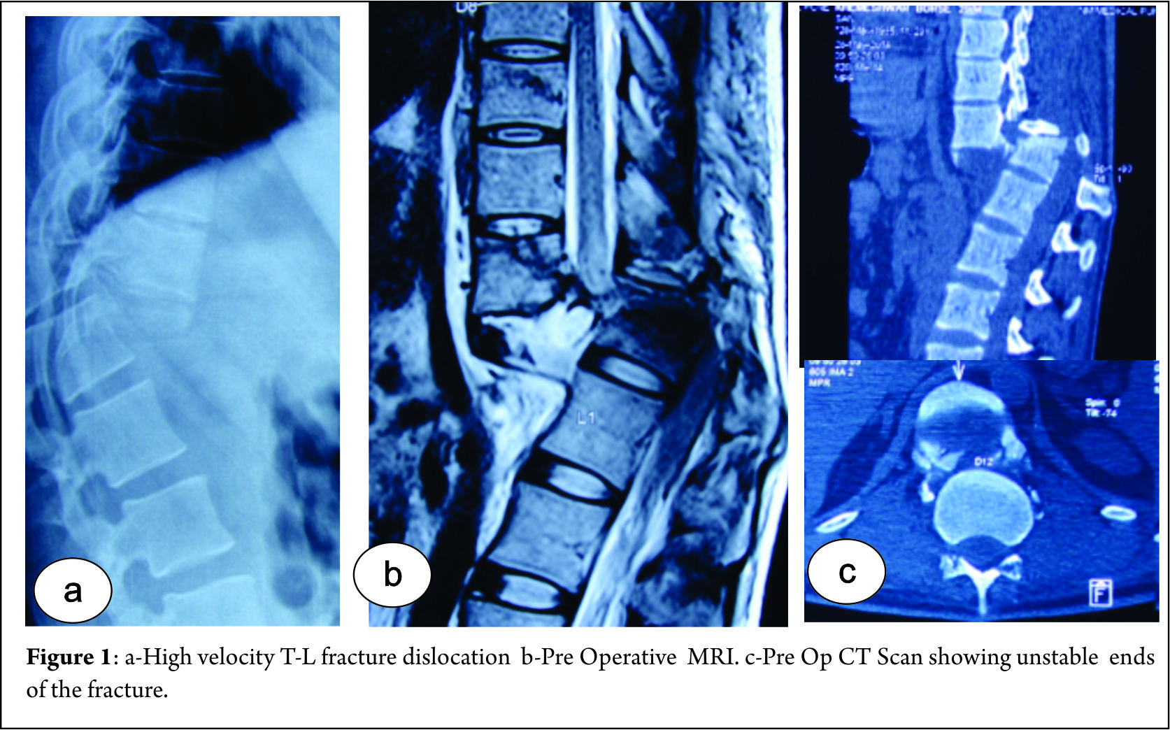
Fracture dislocations of thoracic and lumbar vertebrae are commonly encountered and are a result of high velocity trauma (Fig 1) [1] . Surgical treatment poses challenge in terms of safe and effective reduction maneuvers. Various techniques have been diversely described in literature for reducing highly unstable thoracolumbar fractures. We describe a novel reduction maneuver which is slow, steady and graded technique using four parallel rods and vice grips. We believe that this technique is easy to perform and reproducible, tailor made and maintains good sagittal and coronal balance of spine in such unstable situations. These surgeries are done for mainly early mobilization with a stable spine understanding that the neurology will rarely improve.

Figure 1
Surgical technique:
1) The patient is positioned prone very carefully taking care to prevent further neurological insult. Maintaining the lumbar lordosis can help in partial vertebral reduction.
2) Standard Posterior midline approach taken and exposure done. Utmost care taken at fractured vertebrae level to prevent neural injury.
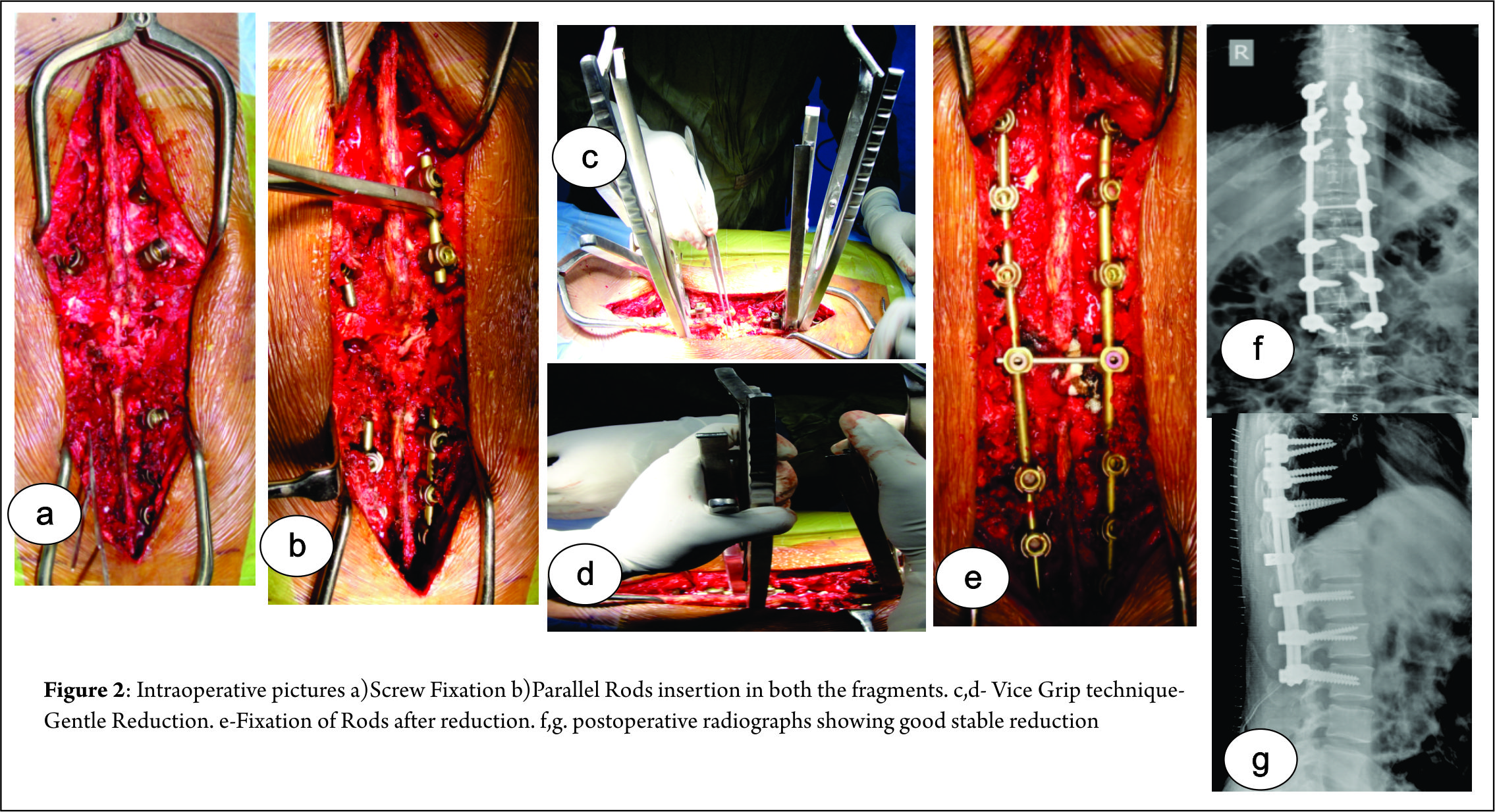
3) Pedicles identified and screws are inserted at two levels above and below the level of dislocation bilaterally (Fig 2a). Role of assistant surgeon is very important in view of giving the counter traction and support the completely shattered spine while inserting pedicle screws. We have seen the shortening of the column because of overlapping of two diseased ends.
4) Two small rods are applied: one below the level of injury and other above (Fig 2b). Once the rods are secured, a three step maneuver is performed. Two rods are held securely by two vice grips and gentle, slow and sustained distraction applied across the vertebrae (Fig 2c). With distraction maintained the surgeon gently pushes the anteriorly displaced proximal column and pulls the distal column (commonly encountered situation where proximal column is anteriorly displaced with respect to distal one) leading to unlocking and disimpaction of dislocated vertebrae. Now opposite forces are applied pulling the proximal column and pushing the distal column to achieve reduction of facet joints bilaterally (Fig 2d). The reduction is checked under fluoroscopy. Sham rod is immediately fixed before the check image.
5) Care should be taken to avoid unnecessary jerky and violent movements. Any bony or soft tissue structures obstructing the reduction should be cleared before attempting reduction.
6) The operating surgeon maintains the reduction and the assistant applies rod on the opposite side and temporarily fixes the vertebral column in this position. Now the temporary rods are removed and another long rod is applied on other side too (Fig 2e).
7) Complete decompression is performed at injured level and neural elements checked for any breach and repaired .Cross links applied.
8) Autografts from laminae and spinous process along with iliac crest bone graft used to enhance fusion.
9) We recommend a CT scan with reconstruction in every case possible along with X rays and MRI scans (Fig 2f,g)

Figure 2

The patient is mobilized as per tolerance with help of brace from second postoperative day in most of the cases.

Discussion
Fracture dislocations of vertebral column are highly devastating injuries and are caused as a result of high velocity trauma. These are commonly seen due to road traffic accidents, fall from height and are often facilitated by risk factors like rheumatoid arthritis etc [1,2]. The mechanism of such injuries varies and is usually a combination of flexion-distraction, translation-shearing or hyperextension injuries [1,3].
Reduction of thoraco-lumbar vertebral fracture dislocations are diversely described in literature. Closed reduction technique using cotrel frame was described initially [4] . However recent trend is shifted towards open reduction which enables us to see neural elements directly and allows adequate decompression. Open reduction techniques described involved direct manual reduction, interspinous lamina spreaders etc [5,6,7]. However they are associated with high failure rates, therefore more aggressive techniques including facetectomies, laminectomies etc are now employed. Our technique described, provides excellent means of reducing highly unstable vertebral dislocations with minimal risk of neural injury. It provides graded, sequential reduction after securing proximal and distal anchorage points (small rods).Although this technique significantly aids in reduction, it should be remembered that it can be difficult at times to replace temporary rods with longer rods.as t is one under vision, it’s easy to be safe while doing this manoevour.
We keep a detailed neurological chart and ASIA impairment scale.

Conclusion
We believe that this technique provides safe and easy way to achieve reduction and maintain sagittal alignment. It is easily reproducible, but we would like to emphasize the importance of a experienced surgeon to prevent additional neural injury while manipulation. Also we would like to mention that this technique is aimed at stabilization for wheelchair mobilization and understanding neurological damage and recovery.This technique helps in avoiding the unnecessary bone cutting/osteotomies as well as shortening procedures and reduces morbidity as it reduces significant time also in difficult reductions.


References 

1. Vialle R, Charosky S, Rillardon L, Levassor N, Court C(2007). Traumatic dislocation of the lumbosacral junction diagnosis, anatomical classification and surgical strategy. Injury 38:169-81.
2. Hauge T, Magnaes B, SkullerudK(1980). Rheumatoid arthritis of the lumbar spine leading to anterior vertebral subluxation and compression of the caudaequina.Scand J Rheumatol 9(4):241-4.
3. Cho Sk, Lenke LG, Hanson D(2006). Traumatic noncontiguousdouble fracture-dislocation of the lumbosacral spine. Spine J 6(5):534-8.
4. Kerboul B, Lefevre C, Le Saout J, Mener G, Miroux D, Roblin L, Courtois B(1986).[Stabilization of thoraco-lumbar spinal fractures using Harrington’s equipment.Study of the evolution of spinal curvatures]. Neurochirurgie 32(5):391-7.
5. Ovejero AMH, Mata SG, Lecea FJM, Zubiri IG, Zubiri LA(2010).L3-L4 Dislocation without Neurological Lesions. Bulletin of the NYU Hospital for Joint Diseases 68(1):60-4.
6. Fazl M, PirouzmandF(2001). Intraoperative reduction of locked facets in the cervical spine by use of a modified interlaminar spreader: technical note. Neurosurgery48:444-5.
7. Lim MR, Mathur S, Vaccaro AR(2009). Open reduction of unilateral and bilateral facet dislocations. In: Vaccaro AR, AlbertTJ (ed). Spine surgery: tricks of the trade. 2nd ed. Thieme,new york;p18-20.


How to Cite this Article: Hadgaonkar S, Khurjekar K, Shah K, Kothari A, Shyam A, Sancheti PA Novel Reduction Manoeuver for Highly Unstable Thoraco-Lumbar Fracture Dislocation:  Technical Note. International Journal of Spine Apr – June 2016;1(1):40-42 .

Photo


(Abstract)      (Full Text HTML)      (Download PDF)


Assessing Frailty in elderly undergoing spine surgery

Volume 1 | Issue 1 | Apr – June 2016 | Page 39|Kunal Shah[1], Manish Kothari[2], Abhay Nene [1]


Authors :Kunal Shah[1], Manish Kothari [2], Abhay Nene[ 1]

[1]     Department of Spine Surgery, Wockhardt Hospital and Medical Research Centre Agripada, Dr Anand Rao Nair Road, Mumbai Central, Mumbai India – 400008.
[2]    Consultant spine surgeon, Suchak hospital, Manchubhai road, Malad east, Mumbai-97.

Address of Correspondence
Dr Kunal Shah
Department of Spine Surgery, Wockhardt Hospital and Medical Research Centre Agripada, Dr Anand Rao Nair Road, Mumbai Central, Mumbai India – 400008.
Email: orthokunal@yahoo.com.


There has been a significant increase in elderly population undergoing spine surgery. This is likely because of improved anesthetic and surgical techniques. Recent literature suggests spine surgery even in older population is safe. With an increase in life expectancy, awareness of recent advances and desire to improve quality of life, many elderly patients opt for surgery. Surgeons are not hesitant to offer surgery in the elderly in view of improvements in the perioperative care. Despite advances in medical care, and being medically “fit”, a small subset of patients suffer from adverse postoperative events. There is a lacuna to identify these high risk individuals and predict postoperative adverse outcome. Older age and pre-existing comorbidities are known predictor of postoperative adverse outcome. However, these are not accurate. Recently Frailty assessment has emerged as independent predictor in various surgical procedures of postoperative morbidity, length of ICU stay, length of institutional stay and mortality [1]. In spine surgery we are often faced with clinical dilemma in terms of offering spine surgery in few scenarios like in elderly patients with degenerative pathology severely hampering daily activities and quality of life or in elderly patients with tuberculous spondylodiscitis where chemotherapy is the primary line of management, but presenting with neurological involvement and/or instability warranting surgery etc. In such situations many questions arise like whether to operate or not and let patient live with disability? If we operate then what is the risk and is there any method of predicting it? Therefore careful assessment of risk –benefit ratio in terms of morbidity/mortality post-surgery should be adequately done for preoperative counselling and predicting outcome. Frailty scoring can be useful in terms of quantifying preoperative risk factors and identify potential modifiable risk factors in such settings. Patel et al [2] concluded that modified frailty index is helpful in predicting mortality in patients with fracture neck of femur. Frailty refers to a condition or syndrome characterized by multisystem decrease in reserve capacity. It encompasses complex issue of disability, comorbidity, cachexia and sarcopenia. Frailty has proven to be associated with mortality and morbidity in short term and long term basis. The prevalence of frailty in patients of all ages presenting for surgical procedures is quoted between 4.1% and 50.3%. Frailty scoring has been successfully used in gastrosurgery, hepatic surgery, cardiovascular surgery, neck of femur fracture surgery etc. Its use in spine surgery is not validated [1,3]. The ideal tool for measuring frailty should serve two purposes: quantify risk and point out modifiable risk factors. The measurement of frailty has been done with various tools like grip strength, gait speed, Edmonton frail scale, comprehensive assessment of frailty score etc. Modified frailty index developed from Canadian study of health and aging has been successfully used in orthopedic setting. Modified frailty index [2] consists of 19 clinical deficits based on 70 potential deficits described in Canadian study of health and aging. It includes parameters like cerebrovascular events, Cardiac diseases, congestive heart failure, psychosis, depression, cognition, diabetes, syncope, recurrent falls, ambulation, seizures, malignancy, Parkinson’s disease, urinary incontinence, renal disease, respiratory disease, decubitus ulcers and myocardial infarction. This is fairly detailed information about most important body systems, however it may be too detailed for spine surgery patients. However similar but more concise scoring system is required to identify high risk individuals in spine surgery.
There is variable amount of physiological capacity in elderly presenting for spine surgery. Therefore predictive models like frailty scoring appear to be useful in predicting morbidity and mortality. Such scoring can potentially help in better preoperative counselling, aggressive physiotherapy, more frequent visits by primary care physician, better optimization of patient prior surgery and affect economics in treatment.

References
1. Partridge JS, Harari D, Dhesi JK. Frailty in the older surgical patient: a review. Age Ageing. 2012;41(2):142-7.
2) Patel KV, Brennan KL, Brennan ML, Jupiter DC, Shar A, Davis ML. Association of a modified frailty index with mortality after femoral neck fracture in patients aged 60 years and older. Clin Orthop Relat Res. 2014;472(3):1010-7.
3) Makary MA, Segev DL, Pronovost PJ et al. Frailty as a predictor of surgical outcomes in older patients. J Am Coll Surg 2010; 210: 901–8.


How to Cite this Article:  Shah K, Kothari M, Nene A. Assessing Frailty in elderly undergoing spine surgery. International Journal of Spine Apr – June 2016;1(1):39 .

Photo


(Abstract)      (Full Text HTML)      (Download PDF)


Spine Surgery In India The Past, The Present & The Future

IJS COVER thumb

Vol 2 | Issue 1 |  April – June 2016 | page:3-4 | Dr. Ram Chaddha


Author: Dr. Ram Chaddha [1]

[1] President – ‘Association Of Spine Surgeons of India – 2015-17’
President – ‘Bombay Orthopaedic Society – 2015-16’
Council Member – ‘APOA/ APSS – 2016-17’
Prof. of Orthopaedics – K.J. Somaiya Medical College, Mumbai
Consultant – Lilavati, Wockhardt, Saifee Hospitals – Mumbai


Spine Surgery In India The Past, The Present & The Future

“It is the best of times….
It is the worst of times….”

PAST: As I look back over the past three decades, I can but reminisce over how much things have changed and yet how little things have changed in “Spine Surgery” in our country. The only permanent thing in life is “change”, but has this “change” really changed anything?
As a young Orthopod in training, from 1986-88, I recollect the despair, disgust and desperation of holding a retractor, almost falling asleep and into the surgical wound, as my most respected senior surgeon spent hours battling his way through an “ Antero-lateral decompression” for a tuberculosis of the mid-thoracic spine. I recollect having sworn, that I would never do this sub-specialty in my future. Probably this was just the “caterpillar” being challenged and the transformed “butterfly” became a ‘Spine Surgeon”.

PRESENT: The long drawn procedures with low illumination, poor magnification and less optimized anesthesia, have been replaced by much better intra-operative conditions, making the “objective “ surgical environment better, but…
Have outcomes really improved? Have the fears and reservations of patients reduced? Have surgical indications become more rationalized? – The answers to the three aforementioned questions are – YES & NO. This is the paradox that the “Spine Surgeons” face today.

Have outcomes really improved?
Both Orthopaedic & Neurosurgery trained Spine Surgeons practice this unique sub-specialty, which truthfully should be a team approach, so well conceptualized and executed at very few centres in our country. Conventional long incisions have been challenged by “minimal access” approaches, supposedly reducing the morbidity. However, have we truly found a solution that optimally and adequately does the basics of spine surgery – decompression/ stabilization/ reconstruction/correction? – YES & NO.

Have the fears and reservations of patients reduced?
Much as it may appear that patients are consenting easily to “spine surgery”, the fears and reservations still remain. The image of the anecdotal and at times factual post-operative paraplegic with incontinent sphincters, looms large on the mind of the lesser informed. The Internet savvy, better informed yet lesser learned, challenge the wits of the treating surgeon putting him on the defensive making him medico-legally tentative and compromising decisions at times. This aspect of the “patient & family psyche” needs significant re-education and proper direction. Have we found an “S.O.P” (Standard Operating Procedure) for this? – YES & NO.
Have surgical indications become more rationalized?
Much as surgeons may feel convinced that all surgical indications are algorithmic and based on evidence-based decisions, there is the humongous pressure of the trade and industry which seems to be driving the evidence and channelizing or polarizing techniques, approaches and expensive consumables making “gold standard” healthcare out of the reach of the common man. The “eye does not see, what the mind does not know”. There are large caveats in our practice that remain mystical. The degenerated disc, the adjacent segment disease and of course the clinico-radiological mismatch very often hound the wits of even the most brilliant minds. Are we reaching the “correct answer” or is it still a “perception of truth”? – YES & NO.

FUTURE: I have a dream, yet unfulfilled which is futuristic.
The surgical decision should be taken only when all other measures seem inadequate. Patient evaluation, counseling, consenting with risk-benefit stratification made more elaborate, critical and standardized. A complete “SWOT – Strength-Weakness-Opportunity-Threat Analysis” of the procedure offered be available for and understood by the patient and the family. No fine print. NO E & O – Errors and Omissions – Excuse. NO* – *CONDITIONS APPLY.
The thinking process of the patient and the treating surgeon needs to change. The patient needs to respect the “intent to treat” of the surgeon and not look at him as a service provider and himself as a consumer. The “Spine Surgeon” should be more realistic in his commitment of post-operative result and be “Godly” not “God”.
The “Make In India” initiative should look at local and regional solutions to “Spine Surgeries”, both as regards decision-making and surgical options.

I strongly believe in “First, do no harm …” – commonly attributed to Hippocrates of Kos, c. 460 BC.
We all need to “pause” and “think”….
– How am I doing?
– What am I doing?
– Where am I going?

It is far less frustrating to have a non-surgical unhappy patient rather than a post-operative unhappy patient….

The author is
Ram Chaddha, M.S. (Orth.)


How to Cite this Article: Ram C. Spine Surgery In India: The Past, the Present & the Future…. International Journal of Spine Apr – June 2016;1(1):3-4.

Dr Ram Chaddha

 Dr Ram Chaddha


(Abstract)      (Full Text HTML)      (Download PDF)