
Vestigial Tail Associated to Spinal Lipomas: Report of Seven Cases
Volume 8 | Issue 1 | January-June 2023 | Page: 07-10 | Arce-Lozoya Mayra Alejandra, Ramírez-Reyes Alma Griselda, Arredondo-Navarro Luis Ángel, Rajendra Sakhrekar
DOI: https://doi.org/10.13107/ijs.2023.v08.i01.45
Submitted: 09/04/2023; Reviewed: 26/04/2023; Accepted: 09/05/2023; Published: 10/06/2023
Authors: Arce-Lozoya Mayra Alejandra [1], Ramírez-Reyes Alma Griselda [1], Arredondo-Navarro Luis Ángel [2], Rajendra Sakhrekar [3]
[1] Department of Pediatric Neurosurgery, UMAE Hospital, Siglo XXI, CdMx, México.
[2] Department of Pediatric Neurosurgery, Hospital Civil Fray Antonio Alcalde, Guadalajara, Jalisco, México.
[3] Department of Orthopaedics, The Hospital For Sick Children, Toronto, Canada.
Address of Correspondence
Dr. Arce-Lozoya Mayra Alejandra
Department of Pediatric Neurosurgery, UMAE Hospital, Siglo XXI, CdMx, México.
E-mail: mayra.arce.lozoya@gmail.com
Abstract
Objective: Vestigial tail is a rare form of cutaneous stigmata often related to occult spinal dysraphism. We present 7 cases of vestigial tail associated to tethered cord and spinal lipomas in the Pediatric Neurosurgery Service of the Hospital Civil de Guadalajara.
Methods: In this retrospective study from 2017-2022 we reviewed the spinal dysraphism data base of the Hospital de Guadalajara and included the cases of vestigial tails.
Results: 7 patients, 5 female and 2 males with vestigial tails were included. Six different morphologies of tails are described so as their location and relationship to occult spinal dysraphism. All the patients received surgical treatment. Complete resection of the cutaneous stigmata and cord detethering were performed in four cases related to fatty filum terminale and the remaining three of the patients required radical resection of associated complex conus medullaris lipomas. All the surgical procedures were performed with microsurgical techniques and neurophysiological monitoring.
Conclusions: The different classifications include two main forms of tails, true tail and false tail. Most true tails represent only a cosmetic issue, while the false ones are often in close relationship with spinal dysraphism. Considering the risk of underlying malformations associated with false human tails, the preoperative assessment should include a thorough neurological examination, and a spine magnetic resonance imaging. We present the association of several forms of spinal lipomas related to vestigial tails.
Keywords: Vestigial tail, Tethered cord, Spinal lipoma, Pseudo tail, Occult spinal dysraphism.
Introduction
Occult spinal dysraphism (OSD) is commonly characterized by skin-covered lesions mainly localized in the lumbosacral area. As skin and nervous tissue are of ectodermal origin, anomalies of both may occur at the same time [1]. Vestigial tails (VT) are rare and heterogenic forms of cutaneous stigmata and might be related to different forms of tethered cord (TC) and OSD.
Most of the knowledge related to VT comes from case reports, thus large series are unavailable. Many descriptions and classifications have merged since Bartles first described it in 1884. Harrison in 1901 made the first differentiation amongst the true and the false tail, naming the nonbony lesion as “caudal appendage”. Since then, various authors presented their different classifications until Dao and Netsky in 1984, published their classical description of “true human tail” and “pseudo tail” which is grossly still in use nowadays [2, 3].
Based on these descriptions, “true tails” include vertebrae, although their embryological origin is different than the false or vestigial tails. VT are different forms of appendages covered by skin and mainly composed of mesenchymatous tissues including fibrous, adipose, and sometimes striated muscle. Nerves can be observed in pathological specimens. These VT are associated with OSD in some cases [3] and represent a major risk to develop a TC syndrome while the child grows up [4].
The report of different forms of spinal lipomas and cord TC in association to VT is not often found in the current literature, we present though 7 cases of VT associated to a different variety of spinal lipomas and TC in the Pediatric Neurosurgery Service of the Hospital Civil de Guadalajara, México.
This case report has been reported in line with the SCARE Criteria [10].
Material and Methods
We reviewed the spinal dysraphism data base of The Hospital Civil de Guadalajara from 2017 to 2022 and selected 7 cases of vestigial tail: 5 female and 2 males. The archives included clinical photos and study images along with the clinical charts. An informed consent was achieved from each of the cases and the identity of the patients was preserved under the regulations of good medical practices. A Magnetic Resonance Imaging assessment was performed in each of the patients. All the patients were operated on for resection of the VT and for the different OSD under microsurgical techniques and neurophysiological monitoring. The morphology and location of the VT were classified according to Tojima and Yamada classification 2020 [2].
Results
Seven patients with several forms of vestigial tails were included and analyzed. Demographic data, morphology and location of the tail, clinical findings, type of surgical procedure, type of dysraphism associated and follow up are shown in table 1.
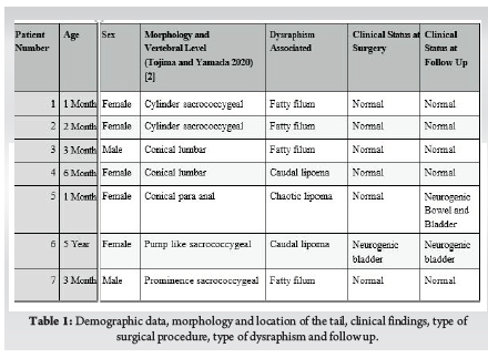
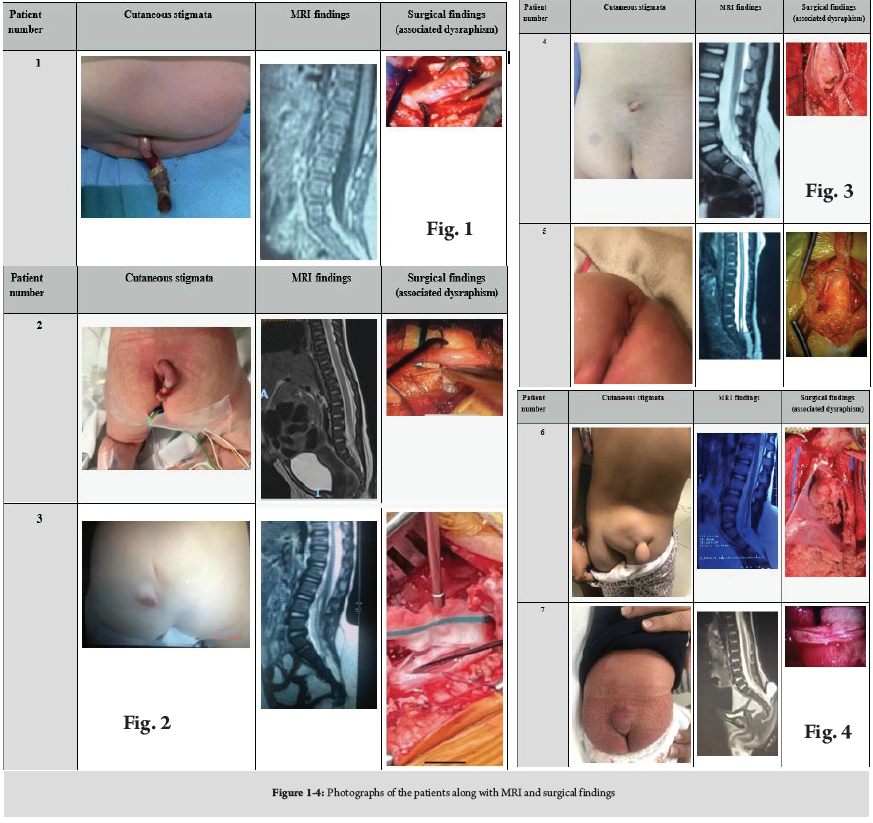
A total of 7 patients were included, 5 female and 2 males; the age range was 1 month old to 5 years old, with a median of 3 months. The aspect of cutaneous stigmata was remarkably heterogeneous varying from tail shaped lesions (cases 1 and 2) to appendicular lesions (cases 3, 4, 5, 6, 7) All of the cases had a preoperative MRI, the lower conus medullaris position below the L2-L3 transition was common in all the cases, a fatty filum terminale was observed in 4 cases (patients 1, 2, 3 and 7), and lipomas of the conus medullaris in three, 1 transitional chaotic lipoma (patient 5) and two caudal lipomas (patients 4 and 6). The clinical preoperative status was normal in almost all the cases, patient 6 had neurogenic bladder though. All the patients were operated on under microsurgical technique and strict neurophysiologic monitoring. Findings during surgery corresponded to preoperative MRI. The postoperative clinical follow up remain unchanged in patients 1, 2, 3, 4, 6, 7 and patient 5 developed neurogenic bladder during her growth (Fig. 1-4).
Discussion
VT are rare embryonic anomalies occurring in the lumbar, sacrococcygeal and peri anal regions. Covered in skin, these lesions are mostly pseudo tails and have an enhanced relevance due to its association to several forms of OSD. On the other hand, the true tails include bone and resemble more to those present in other non-human vertebrates [2, 9]. VT probably occur during the 5th and 6th gestational week and this represent their common association to OSD and TC.
Many attempts to classify this anomaly have being used throughout the times and are cited in most of the papers referring these cases [2, 8]. Most of classifications of the so called “true” or “false” tails are based on the presence of bone, calling “true tails” to those bonny appendages. Although the reports of true tails composed of bony elements are extremely rare (counting just a few cases described) may still causing confusion now to establish a fine description [11]. It is important to establish that VT or pseudo tails are more often related to an OSD and must be considered under the spectrum of cutaneous stigmata requiring a more accurate screening of the patients. Thus, in the strict sense, the description of a true tail must not be confused with a VT in terms of treatment, assessment and follow up [11].
We found the classification made by Tojima and Yamada in 2020 very useful. They described three main anatomic locations and six shapes of the vestigial tails. In our point of view this is a simple and accurate way to classify the lesions [2, 3].
In this paper we present the association of different spinal lipomas in all the cases. Four filar lipomas and three lipomas of the conus medullaris, according to the several classifications for spinal lipomas [5] Spinal lipomas are not simple lesions and require a vast experience of the surgeon to treat them, especially in cases of those of the conus medullaris [6].
We found no reports of transitional chaotic lipomas associated to peri anal vestigial tails, as we found in the case number five; so, this might be considered in the assessment of patients with these anomalies. The development of neurogenic bladder in this case could be related more to the type of spinal lipoma she presented, rather than the result of the surgical procedure. Chaotic lipomas are associated to an uncertain prognosis no matter the moment the surgery is performed [6].
The four filar lipomas were completely excised, along with the correction of the cutaneous tail. In the more complex conus medullaris lipoma cases, an aggressive radical resection was attempted to remove most of the lipoma, in two cases the placode neurulation was feasible.
The seven cases we present are of utmost interest because we encountered a very strong relationship of VT to different forms of spinal lipomas. If we referred to the embryonic classification of these lesions, Morota et al. published a brand-new classification of spinal lipomas according to embryonic stage [5]. All our patients presented some different forms of spinal lipomas, 4 fatty filum terminale, 2 caudal lipomas and 1 chaotic lipoma. This finding probably can lead us to realize if there is a relationship between the cutaneous stigmata and the subjacent OSD according to the embryonic stage, and may be important to encourage the need for a complete screening of the spinal cord rather than the simple cosmetic resection of the VT.
We emphasize the importance of the risk of underlying spinal lipomas and tethered cord associated to these vestigial tails. Several other reports have mentioned the associations of dermal sinus and other forms of OSD [7]. Dermal sinus tract is as well a very concerning OSD due to the association of spinal cord dermoid cysts.
The presence of a VT is a matter of concern for the parents, so they might feel under pressure for their child to be operated on as soon as possible. The timing is important for removing the VT and the decision to treat the underlying OSD must be as important as well, thus a thorough assessment since the initial visual diagnosis, must be completed with the appropriate studies, in order to establish the definitive treatment.
The correct recognition of an OSD and its prompt treatment, could be of benefit in preventing future neurologic, orthopaedic or urologic disorders; nevertheless, the early treatment of asymptomatic patients with tethered cord is still on debate and further studies are needed.
Conclusions
1. Most of true tails represent only a cosmetic issue
2. Pseudo-tails are often related to spinal dysraphism.
3. Considering the risk of underlying malformations associated with human tails, the preoperative assessment should include a thorough neurological exam, plain radiographs to clear spinal defects and a spine magnetic resonance imaging.
4. Surgery in these cases should be focused in untethering the cord and the esthetical correction of the cutaneous stigmata.
References
1. Guggisberg D et al. Skin markers of occult spinal dysraphism in children. A review of 54 cases. Arch Dermatol 2004;140:1109-115.
2. Tojima S, Yamada S. Classification of the “human tail”: Correlation between position, associated anomalies, and causes. April 2020 Pages 929- 942.
3. Rueda J et al, Human tail in a newborn. Journal of Pediatric Surgery 2022. 102098.
4. Hertzler DA, De Powell JJ, Stevenson CB, Mangano FT. Tethered cord syndrome: a review of the literature from embryology to adult presentation. Neurosurg Focus 29(1):E1, 2010.
5. Morota N, Ihara S, and Ogiwara H. New classification of spinal lipomas based on embryonic stage J Neurosurg Pediatr 19:428–439, 2017.
6. Pang D. Management of complex spinal cord lipomas: a new perspective. J Korean Neurosurg Soc. 2020 May; 63(3) 279-313.
7. Mukhopadhyay B, et. Al. Spectrum of human tails: A report of six cases J Indian Assoc Pediatr Surg. 2012 Jan-Mar; 23–25.
8. Wilkinson CC, Boylan A. Proposed caudal appendage classification system; spinal cord tethering associated with sacrococcygeal eversion. In Child’s Nervous System Ausgabe 2017.
9. Turk CC, Kara NN, Bacanli A. The human tail: a simple skin appendage or cutaneous stigma of an anomaly?. Turk Neurosurg. 2016;26:140-5.
10. Agha RA, Franchi T, Sohrabi C, Mathew G, Kerwan A. The SCARE 2020 Guideline: Updating Consensus Surgical CAse REport (SCARE) Guidelines .International Journal of Surgery. https://doi.org/10.1016/j.ijsu.2020.10.034.
11. Pillai MK, Nair ST. A true human tail in a neonate. Case report and literature review. Sultan Qaboos University Med J, February 2017, Vol. 17, Iss. 1, pp. e109–111.
| How to Cite this Article: Arce-Lozoya M A, Ramírez-Reyes A G, Arredondo-Navarro L Á, Sakhrekar R | Vestigial Tail Associated to Spinal Lipomas: Report of Seven Cases | International Journal of Spine | January-June 2023; 8(1): 07-10. |

